Simulations Reveal Supercoiled DNA Is Complex, Wiggly
Just about everyone is familiar with the double helix, the shape of DNA as formed by a pair of molecule strands. The structure was first discovered by Francis Crick and James Watson, and has since become an iconic image found scattered throughout modern artwork, movies, and more. Things get tricky when it comes to full long DNA strands coiled up inside of cells, though, and a team of researchers have tasked themselves with unraveling some of the mysteries.
The researchers, whose efforts were recently detailed in a report in Nature Communications, have set about imaging DNA shapes using a strong microscopy technique. The resulting images were then analyzed via simulations run on a University of Leeds supercomputer.
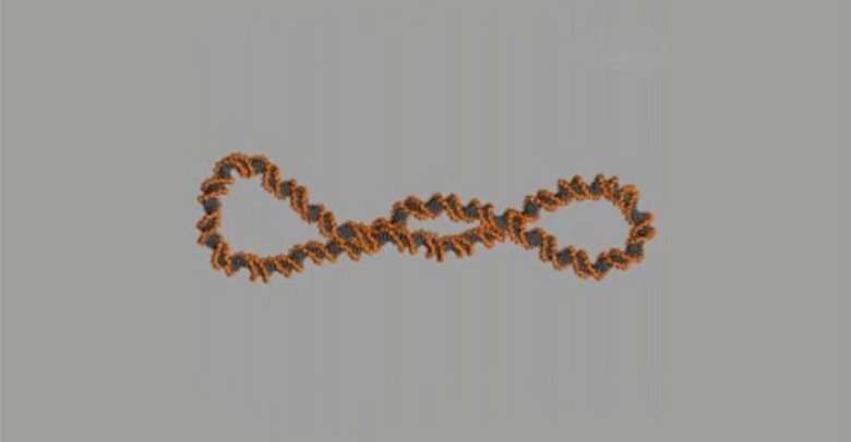
The simulations reveal that DNA is wiggly, for one thing — it constantly shifts and morphs its shape inside of the cells. This makes it far more complex than the popularized double helix DNA. All sorts of shapes can result, everything from "8" shapes to things described like handcuffs or sewing needles.
University of Leeds' Dr. Sarah Harris elaborated on the differences, saying:
When Watson and Crick described the DNA double helix, they were looking at a tiny part of a real genome, only about one turn of the double helix. This is about 12 DNA 'base pairs', which are the building blocks of DNA that form the rungs of the helical ladder. Our study looks at DNA on a somewhat grander scale – several hundreds of base pairs – and even this relatively modest increase in size reveals a whole new richness in the behavior of the DNA molecule.
The work is important, as expanding knowledge of what DNA looks like will ultimately aid in developing new cancer treatments and medication that are more effective than current offerings.
SOURCE: EurekAlert