MIT Microscopy Technique Improves Deep-Tissue Imaging
Scientists at MIT have developed a new microscopy technique that can make finer images of deeper tissue more quickly. The new technique offers the opportunity to obtain high-resolution images of blood vessels and neurons within the brain. Often, to create high-resolution 3D images of tissues such as brain tissue, scientists use two-photon microscopy.That technique involves aiming a high-intensity laser at the specimen to induce fluorescence excitation. The problem is that scanning deep within the brain using that technique can be difficult because light scatters off tissue as it goes deeper, resulting in blurry images. Two-photon imaging is also very time-consuming, requiring exciting individual pixels one at a time. Researchers from MIT and Harvard University developed a modified version of two-photon imaging to image deeper within tissue while performing the imaging much more quickly than the current method.
The team believes their new imaging could allow scientists to obtain high-resolution images of blood vessels and individual neurons in the brain more quickly. The team modified the laser beam coming into the tissue, allowing them to go deeper and perform finer imaging than past techniques. MIT wanted to develop a method allowing the imaging of a large tissue sample at one time while maintaining the high-resolution offered by point-by-point scanning. The researchers came up with a way to manipulate light that shines onto the sample.
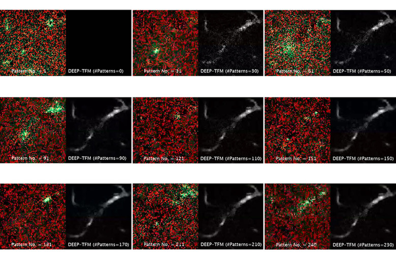
Their breakthrough uses a form of wide-field microscopy that shines a plane of light onto the tissue, but the amplitude of the light is modified, allowing the researchers to turn each pixel on or off at different times. Some pixels are lit while nearby pixels remain dark, creating a predesigned pattern that can be detected in the light scattered by the tissue. After obtaining raw images, each pixel is reconstructed using a computer algorithm created by the researchers.

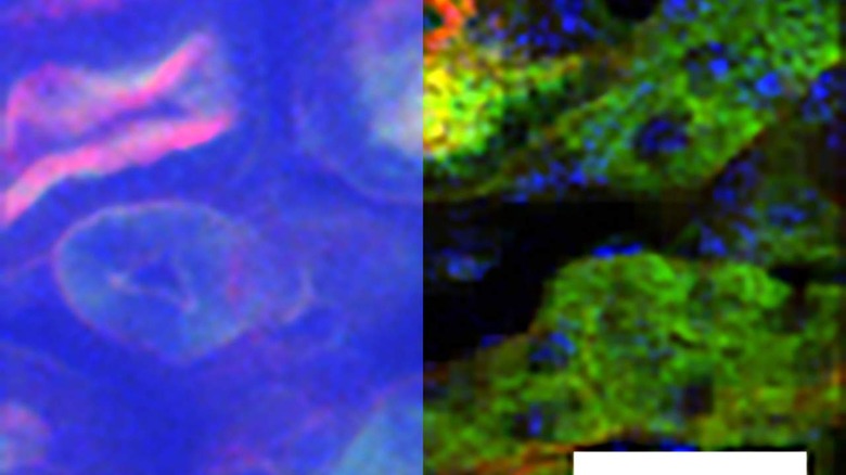
The technique allowed imaging about 200 microns deep into slices of muscle and kidney tissue. It was also able to image about 300 microns into the brains of mice. That is twice as deep as possible without using the patterned excitation and computer algorithm for reconstructing the images. The technique is also able to generate images between 100 to 1,000 times faster than conventional two-photon microscopy.
