Deep-Brain Stimulation Leads To Milestone Discovery About Memories
Researchers from UT Southwestern made a discovery that holds the potential to help improve treatment for people with diseases such as Alzheimer's, schizophrenia, and those suffering from traumatic brain injury. During the research, the team identified characteristics of over 100 memory-sensitive neurons they believe play a role in how the brain recalls memories. Researcher Bradley Lega, M.D., believes the discovery could lead to new deep brain stimulation therapies for various brain injuries and diseases.
Human memory breakthrough
Among the most significant findings in the research was that neuronal firing happens with different timing than other brain activity when the brain is recalling memories. The timing difference is called "phase offset," which is the first time the phenomenon has been reported in humans. Phase offset was previously seen only in rodent studies. The team believes their research shows how the brain can recall an event while keeping track of whether the memory is something new or something previously experienced.
Dr. Lega says the research is some of the best evidence showing how the brain works when recalling old memories compared to forming new memories. The study identified 103 memory-sensitive neurons in the hippocampus and entorhinal cortex that have increased activity when memory encoding occurs. When that memory is recalled, the same pattern of activity occurs again when study participants attempt to recall the same memories, particularly when the memories are highly detailed.

Of particular interest is activity in the hippocampus, which could have relevance in schizophrenia, a disease known for hippocampal dysfunction (via Nature). In schizophrenia, the hippocampal dysfunction leads to the sufferer's inability to distinguish between memories and hallucinations or delusions. Carol Tamminga, M.D, an expert on schizophrenia, says that in people with psychotic illnesses, hallucinations and delusions are actual memories.
Tamminga said that in people with psychotic illnesses, hallucinations and delusions are processed through neural memory systems just as normal memories are, despite the corruption in hallucinations and delusions. Tamminga says it is important to understand how the "phase offset" mechanism might be used to modify corrupted memories.

An opportunity to learn more about how people form memories was presented during a study conducted on people with epilepsy. That study implanted electrodes into the epileptic's brain to map seizures. Lega and his team believed the same electrodes could be used to identify neurons involved in memory.
The epilepsy study involved 27 patients. Lega and his team used the data to identify neurons involved in memory by having the patients participate in memory tasks. However, the team is clear that the data gathered doesn't conclusively prove anything, instead adding credibility to a memory model called Separate Phases at Encoding And Retrieval (SPEAR) developed from rodent studies.
Lega says it's one thing to have a rodent model and another thing to have evidence showing the model is occurring in humans. The SPEAR model predicts the "phase offset" discovered in the new study.
Other neuron studies
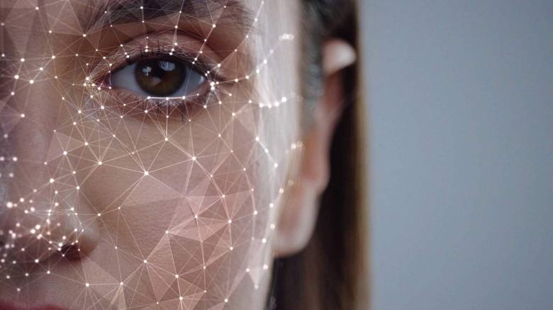
Substantial amounts of research focus on the brain's neurons to learn more about how they function and how they are responsible for memory. In July 2021, researchers identified neurons in the brain responsible for remembering faces (via Science Daily). The team attempted to identify the neurons responsible for the recognition people feel when they see a familiar face.
In that research, the team dubbed the neuron the "grandmother neuron" due to the flash of recognition when people see very familiar faces. The researchers described that neuron as the crossroads of sensory perception and memory potentially responsible for allowing humans to prioritize an important face over others. The team identified a class of neurons located in the temporal pole region of the brain in their research.
Interestingly, while it was typically thought of as a single cell, it turned out to be a population of cells that work together to remember faces. The neurons in the temporal pole region of the brain are highly selective and appear to remember faces the subject has seen before more strongly than unfamiliar faces. In addition, researchers discovered that the neurons could quickly determine between known and unknown faces immediately upon seeing the image.
The research determined that the neurons responded three times more strongly to familiar faces than unfamiliar faces. Project researchers said the ability of the cells to respond to familiar stimuli indicates a change in the brain as a result of past encounters.