Researchers Create The First 3D Map Of Heart Neurons
The research team from Thomas Jefferson University has completed the first 3D map of neurons in the human heart. The group says that the information reveals foundational insight into the role of neurons and heart attacks and other cardiac conditions. The team says that while the brain is the primary controller of the heart, the heart has its own "little brain," called the intra-cardiac nervous system (ICN). The ICN supports heart health and can protect cardiac muscle during a heart attack, but it's not clear exactly how it accomplishes these roles.
The ICN is poorly understood, and scientists didn't know where they were located in the heart, how they were connected, and what their molecular properties were. Scientists at the University have now been able to answer those questions in detail. The researchers worked with scientists from different research groups and industry partners to create a dual-approach pipeline.
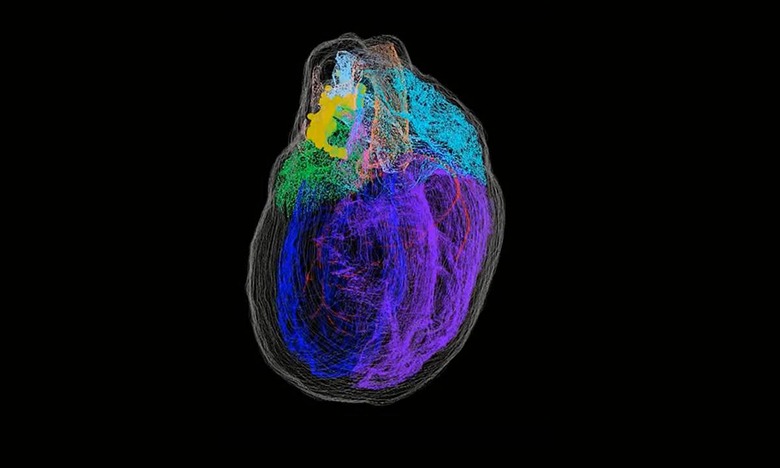
One approach involved a new imaging technique called Knife-Edge Scanning Microscopy that allows the researchers to build a precise three model of the entire rodent heart. The study marked the first use of the technology for cardiac research. The second approach used a technique called laser capture microdissection to sample single neurons for gene expression analysis and precisely map the individual positions within the 3D structure of the heart.
The 3D map created revealed previously unknown complexity of the ICN. Researchers found that the neurons that make up the ICN are found in a coherent band of clusters on the base (top) of the heart where the heart's veins and arteries enter and leave. The ICN also extended down the length of the left atrium to the back of the heart. They're also positioned close to certain key heart structures like the sinoatrial node.
The team compared male and female rat hearts and found that there are sex-specific differences in the way the neurons were organized both spatially and by gene expression. Scientists say that since they know where the neurons are located in relation to heart structures are now able to ask questions like if stimulating one location or selectively stimulating specific neurons make a difference in heart function. The team says they've "created the foundation for an endless possibility of future studies."