MIT Researchers Convert MRI Scans Into 3D Printed Hearts
Some of the challenge for surgeons in preparing for a surgery is getting a good idea of what the area they will be working with looks like. Surgeons use lots of tools to do this such as MRI, x-ray, and other methods to see inside the human body before they cut. Researchers at MIT and Boston Children's hospital have developed a new way to help surgeons see inside the body of children.
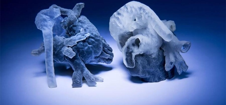
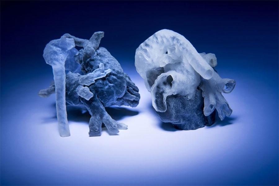
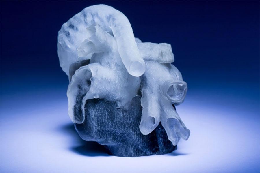
The new system is able to take data from an MRI scan and turn that data into a 3D print of the heart of a child. The process takes unspecified number of hours and once the 3D model is printed, the surgeon can then use the model for surgical planning.

This fall seven cardiac surgeons at Boston's Children's Hospital will be participating in the study to determine if the 3D printed heart models improve surgery outcomes. The new system will be described at the International Conference on Medical Image Computing and Computer Assisted Intervention in October.
For the system to accurately make the models the researchers have a human expert identify and segment a small patch making up one-ninth of the total area of the scans in each cross section. Algorithms then take over to identify the remaining areas. The team says that by segmenting 14 patches the algorithms yield 90% agreement with expert segmentation.
SOURCE: MIT