Scientists develop a new method of creating artificial 3D printed organs
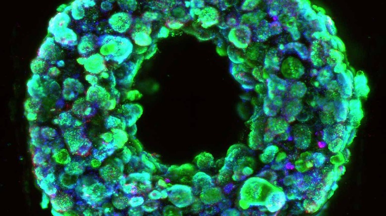
Harvard researchers have created a new method of 3D printing human tissues that could one day lead to 3D printed artificial human organs. The researchers created a new technique called SWIFT (sacrificial writing into functional tissue.) SWIFT overcomes one major hurdle of 3D printing organs by printing vascular channels into living matrices composed of stem cell-derived organ building blocks (OBBs).SWIFT is a 2-step process that begins with forming hundreds of thousands of stem-cell-derived aggregates into a dense, living matrix of OBBs that contain 200 million cells per milliliter. The next step includes creating a vascular network where oxygen and other nutrients can be delivered to the cells embedded within the matrix.
The team says this allows the high cellular density similar to that of human organs and the viscosity of the matrix enabled the printing of a pervasive network of perfusable channels within it to mimic blood vessels to support the organs. The cellular aggregate used in the SWIFT method is derived from adult induced pluripotent stem cells.
At cold temperatures of 0-4 degrees celsius, the matrix has the consistency of mayonnaise. That makes it soft enough to manipulate, but thick enough to hold its shape. When the cold matrix heats to 37 degrees celsius, it stiffens and becomes more solid while the gelatin ink melts and can be washed out.
A network of channels is left behind within the tissue that can be perfused with oxygenated media to nourish the cells. The team was able to print organ-specific tissues with SWIFT that remained viable while tissues printed in other methods died within 12 hours. The team printed and perfused a branching channel architecture matrix of heart derived cells and found the OBBs infused together and the tissue had more contractions that were over 20 times stronger, mimicking features of the human heart.