MIT researchers develop a new, faster 3D brain imaging technique
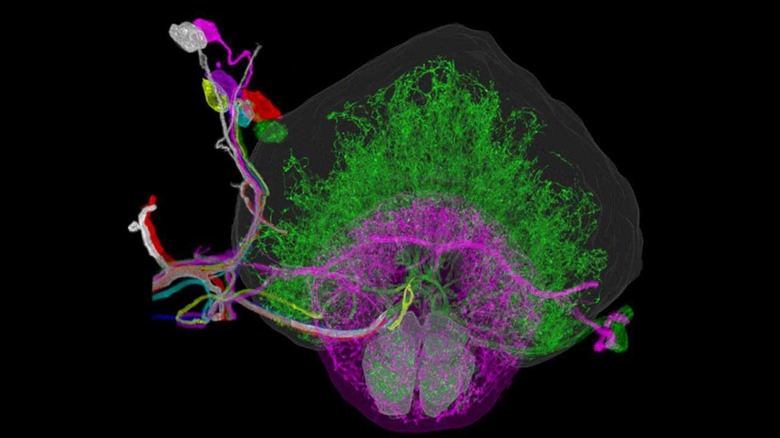
Researchers from MIT have announced that they have developed a new way to image the brain with "unprecedented resolution and speed." The technique allows the location of individual neurons, the tracing of connections between them, and the visualization of organelles inside the neurons over large volumes of brain tissue. The tech combines a method for expanding brain tissue to make it possible to image the tissue at higher resolutions and a 3D microscopy technique called lattice light-sheet microscopy.
The team used the technique to image an entire fruit fly brain and large sections of a mouse brain. They say that the technique is much faster than imaging with previously available technology. One key to the method developed is that it allows the mapping of large-scale circuits within the brain and insight into the individual function of neurons.
"A lot of problems in biology are multiscale," MIT's Edward Boyden says. "Using lattice light-sheet microscopy, along with the expansion microscopy process, we can now image at large scale without losing sight of the nanoscale configuration of biomolecules."
Boyden worked with his team to combine expansion microscopy with lattice light-sheet microscopy for the new study. The latter technology is said to be critical to the new technique and was developed by study co-author Eric Betzig.
Imaging expanded tissue samples generates "huge amounts of data," say the scientists, to the tune of tens of terabytes per sample. This large amount of data meant the team also had to devise parallelized computational image-processing techniques able to break the data into smaller chunks. The smaller chunks were then analyzed and then combined into the whole. Boyden says there are applications beyond neuroscience in cancer and HIV research.